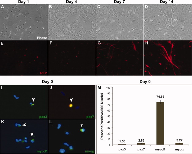
FIGURE 2.
In vitro differentiation of primary myoblasts isolated from α-actin–RFP adult dorsal muscle. (A–D) Phase contrast of zebrafish myogenic progenitor cells differentiating from day 0 to day 14. (E–H) RFP expression of the α-actin promoter indicates myotube formation and myogenic differentiation. (I–L) Immunofluorescent staining of day 0 α-actin-–RFP myoblasts. Note that very few cells express high levels of the α-actin RFP transgene, as it undergoes higher levels of transcriptional expression during myogenic differentiation. Green fluorescent staining and open arrowheads demarcate myogenic markers (pax3, pax7, myod1, and myogenin). (M) Quantification of 500 DAPI-stained (blue) nuclei of the results from day 0 myoblast immunofluorescent staining in (I)–(L). Immunostaining was performed in triplicate. [Color figure can be viewed in the online issue, which is available at wileyonlinelibrary.com.]

