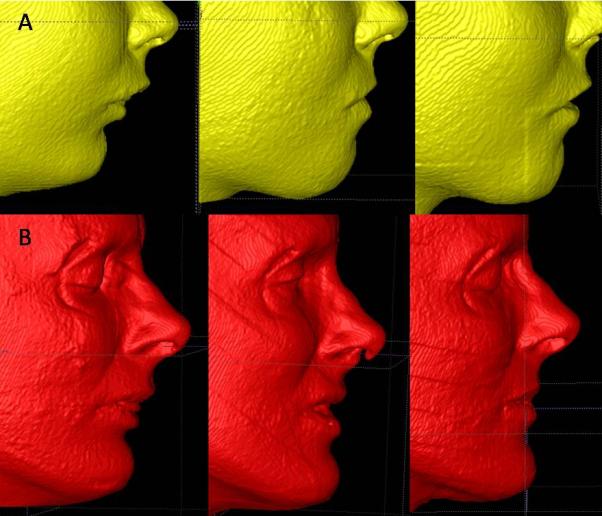
Figure 9.

Example of the three different periods analyzed. Left: images are presurgery. Center: images at splint removal. Right: images at 1 year postsurgery. (A) Patient who had some relapse of the surgery outcomes at 1 year postsurgery. (B) Patients with stable soft tissue results at 1 year postsurgery.
