
Fig. 2.
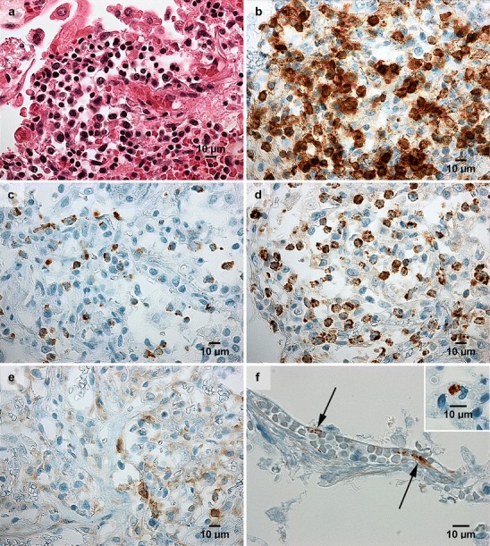
Immunohistochemistry results in lung sections from two fatal cases of hantavirus pulmonary syndrome. The findings were condensed oedematous pulmonary parenchyma with alveolar fibrinous exudate and infiltrates of mononuclear cells (a), whereof a vast majority were CD8+ T lymphocytes (b), and many were holding granules containing granzyme B (c) and T cell restricted antigen-1, TIA-1 (d); this immunophenotype is characteristic of activated cytotoxic T lymphocytes. In contrast, CD4+ helper T lymphocytes (e) were uncommon. Viral antigen was detected in capillary vascular endothelium (f) and in mononuclear cells, here represented by a monocyte (f; inset), using Puumala hantavirus nucleocapsid protein monoclonal antibody (A1C5, Progen Biotechnik GmbH, Heidelberg, Germany). For viral antigen, lung samples from two non-hantavirus patients were used as negative controls (data not shown). Panels (Patient 1 in a–e; original magnification, x 400; and Patient 3 in f; original magnification, x 600) display lung sections from paraffin embedded material. Staining was performed using hematoxylin and eosin (a), and immunoperoxidase technique (b–f)