
Fig. 1.
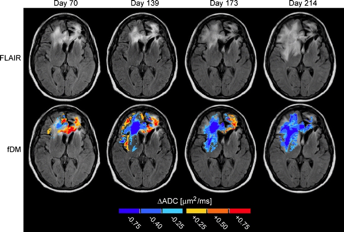
Fluid-attenuated inversion recovery (FLAIR) images and functional diffusion maps (fDMs) in a patient with recurrent glioblastoma treated with bevacizumab and temozolomide. FLAIR images showing gradual spread of nonenhancing tumor (top row). fDMs showing increasing volume of low apparent diffusion coefficient (ADC) (“hypercellular”) regions prior to radiographic failure (bottom row). Image days are with respect to pretreatment baseline (day 0)