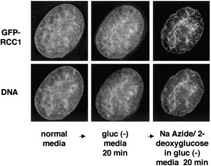
Figure 3.
GFP-RCC1 becomes highly clumped inside the nucleus upon energy depletion. Cells expressing GFP-RCC1 and growing in normal media were assembled in a Bioptechs live-cell chamber with normal media and allowed to equilibrate in a 37°C incubator. After 20 min, the chamber was transferred to a DeltaVision microscopy work station and images were acquired. The media were changed to gluc (-) media, and the same cell was imaged again after 20 min. Finally, the media were changed to gluc (-) media containing sodium azide and deoxyglucose and the same cell was imaged after 20 min in this ATP depletion media. To show the DNA staining in live, unfixed cells, cells were incubated in 1 μg/μl Hoechst 33258 for 20 min before image acquisition. Shown in the figure are single, deconvolved z-sections.

