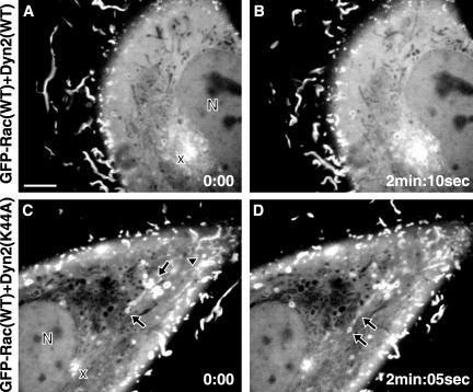
Figure 7.
GFP-Rac(WT) trafficking in the presence of dyn2(WT) and dyn2(K44A). NIH3T3 cells were microinjected with plasmids encoding GFP-Rac(WT) and Dyn2(WT) or Dyn2(K44A). The GFP-Rac(WT) signal was recorded in a time lapse series with a high-resolution spinning disk confocal microscope at the onset of protein expression, before abnormal ruffles became apparent with dyn2(K44A). N, nucleus; x, perinuclear GFP-Rac(WT)-enriched region; arrowhead, GFP-Rac(WT) signal streaks emanating from the zone of membrane ruffles; arrows, tubulo-vesicular structures bearing a membrane-associated GFP-Rac(WT) signal. Time-lapse videos are available as supplemental material. Bar, 5 μm, time as minutes:seconds.

