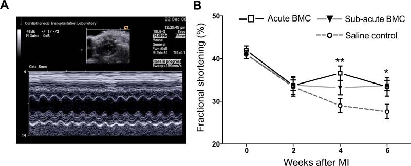
Figure 4. Echocardiographic assessment of cardiac function.
(a) Representative M-mode echocardiogram at the level of the papillary muscle from which left ventricular diameters were measured. (b) Echocardiography revealed a significant preservation of left ventricular fractional shortening at 4 weeks in the Acute BMC group and 6 weeks in both BMC groups compared to saline control animals. No significant difference in cardiac performance was found between Acute and Sub-acute BMC animals. (*P<0.05, **P<0.01, error bars indicate SEM).

