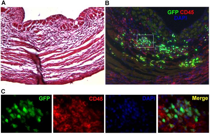
Figure 5. Immunohistochemical analysis of BMC transplanted hearts.
(a) H&E staining of the left ventricular wall shows mononuclear cell infiltrates and scar formation consistent with myocardial infarction. (b) Immunofluorescent staining on a corresponding section reveals an abundant presence of GFP+ BMCs (green) within the infarcted myocardium, which is rich in CD45+ inflammatory cells (red). The majority of the GFP+ cells coexpressed CD45, confirming an inflammatory phenotype. Counterstaining was performed with 4,6-diamidino-2-phenylindole (DAPI, blue). (c) High power views of the selected area (Fig 2B white square) reveal that the vast majority of donor BMCs express CD45 and retain round shapes with large nuclei, representing an inflammatory phenotype.

