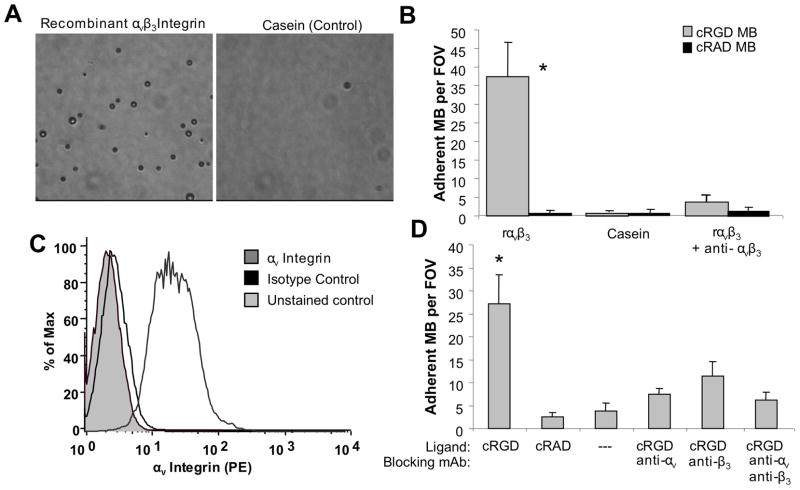
Figure 2.
In vitro microbubble adhesion assays. (A) Characteristic microscopic fields of view for cRGD-MB on recombinant αvβ3 integrin (left) and casein (right) surfaces. (B) Functional adhesion of cRGD and control cRAD microbubbles to recombinant αvβ3 integrin at a wall shear stress of 1.0 dyne/cm2. Adhesion of cRGD-MB was significantly reduced in the presence of an anti- αvβ3 integrin antibody. (C) Flow cytometry results demonstrating αvβ3 integrin expression on bEND.3 murine endothelial cells. (D) Functional adhesion of cRGD and control cRAD microbubbles to bend.3 cells at 1.0 dyn/cm2 in the presence of blocking antibodies against various subunits of αvβ3 integrin. Data are presented as mean +/− standard deviation for n=5 independent experiments. *p<0.01 versus all negative control conditions.

