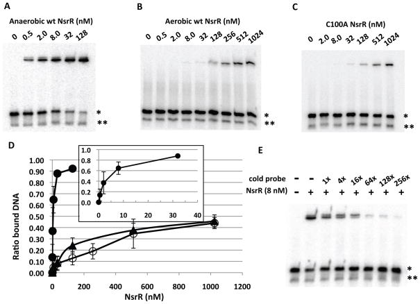
Figure 2.
Binding assay of NsrR to the nasD promoter. The sequence of the nasD probe (−44 to −19) that contains the proposed NsrR-binding site is shown in Figure 1. The radiolabelled probe (0.1 nM) was incubated with increasing concentrations of wild-type NsrR-His6 purified under anaerobic conditions (A), aerobic conditions (B), or with the NsrR (C100A) mutant purified under anaerobic conditions (C). When anaerobically purified NsrR was used, the binding reaction as well as gel electrophoresis was carried out under anaerobic conditions as described in Experimental procedures. A single asterisk shows the double-stranded DNA and a double asterisk shows the single-stranded DNA, unannealed radiolabelled DNA oligonucleotide.
D. ImageJ was used to quantify the ratio of shifted band to total double-stranded probe bands from multiple EMSA experiments (n=6 for A and n=3 for B and C) and the average values are shown with standard deviations. Symbols: closed circles, anaerobically purified NsrR; open circles, aerobically purified NsrR; closed triangles, anaerobically purified NsrR(C100A) mutant protein. The inset shows a binding curve with anaerobic NsrR at concentrations less than 32 nM.
E. Competition assay. Anaerobically purified wild-type NsrR and radiolabelled nasD probe DNA were used with or without excess cold nasD DNA of the corresponding sequence.

