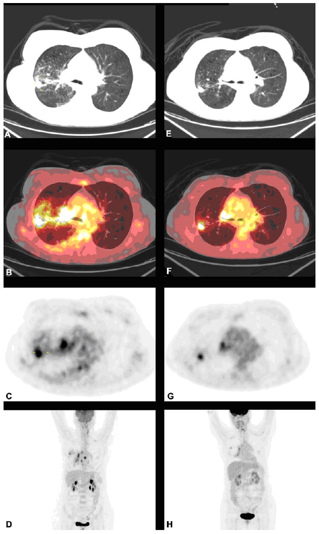
Figure 1.

FDG-PET/CT scans of a 63 year-old female with stage IVA adenocarcinoma of the lung before and after 2 cycles of carboplatin and pemetrexed. Left panel images are pretreatment images (A, B, C and D), while the right panel are posttreatment images (E, F, G and H). The images from top to bottom are CT transaxials (A, E), CT and PET fused transaxials (B, F), PET transaxials (C, G), and maximum intensity projection images (D, H).
