
Figure 3.
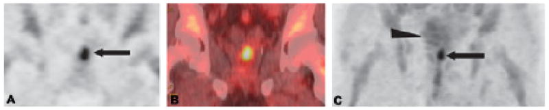
Coronal PET (A) and CT fused (B) anti-18F-FACBC images in a 71-year-old man (restaging) with biopsy-proven prostate bed recurrence extending toward the left seminal vesicle (arrow in A). Maximum-intensity-projection (MIP) image at 20 min (C) demonstrates uptake in the prostate bed (arrow) but little bladder uptake (arrowhead).