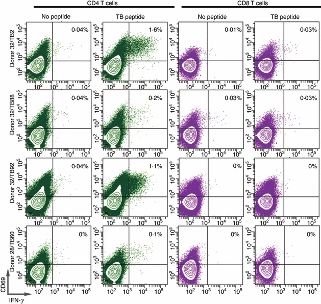
Figure 3.

Intracellular interferon-γ (IFN-γ) production by tuberculosis peptide-stimulated peripheral T cells. Peripheral blood mononuclear cells (PBMC) obtained from Mycobacterium tuberculosis peptide-reactive donors shown in Table 2 were incubated with peptide, for 10 days. Cells were harvested, washed and re-stimulated with the indicated peptide for 4 hr in the presence of Brefeldin A. Cells were then stained by anti-CD4, anti-CD8 and IFN-γ monoclonal antibodies, and analysed by flow cytometry. Notice that the majority of IFN-γ producing cells are in the CD8− CD4+ gated lymphocytes.
