
Figure 2.
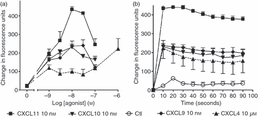
CXCR3 agonists and CXCL4 induce intracellular calcium flux in T cells. Staphylococcus enterotoxin B (SEB)/interleukin-2 (IL-2) activated T cells (day 9–12) were loaded with Fluo-4 AM at 37° for 30 min. Cells were then washed twice in RPMI-1640 medium and aliquoted at 1 × 10⁵ cells/well as described in the Materials and methods and treated with the indicated concentrations of each agonist (a) or with 10 nm of each agonist for the times indicated (b). Changes in fluorescence were measured using multimode plate reader (Fluostar Optima). Peak responses from each stimulation were taken to create a concentration–response curve. Error bars represent mean ± SEM of four experiments using cells from different donors.