
Figure 1.
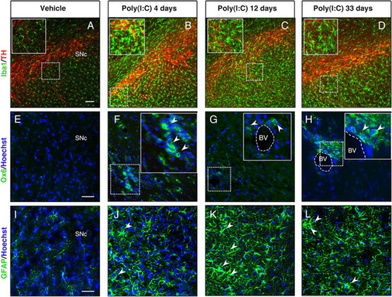
Intranigral injection of poly(I:C) induces microglial and astrocytic activation within the SN. Sprague Dawley rats were injected with vehicle or 20 μg of poly(I:C) into the SN. Brains were isolated at 4, 12, and 33 d after injection, and immunofluorescence microscopy was performed for TH (red), Iba1 (green), MHC class II antigen OX6 (green), and GFAP (green). A–D, Iba1 immunohistochemistry revealed resting highly ramified microglia within the SN in vehicle-injected animals (A). In contrast, animals injected with poly(I:C) at 4, 12, and 33 d after the injection showed activated microglia with shorter and thicker processes and larger cell bodies (insets) (B–D). E–H, MHC-II+ cells (arrowheads) were present in poly(I:C)-injected rats at 4,12 and 33 d after injection. I–L, Astrocytic reaction (arrowheads) was found in the SN at 4, 12, and 33 d after poly(I:C) injection. Cell nuclei were counterstained with Hoechst. Dashed lines in G and H indicate blood vessels. Scale bars: A–D, 100 μm; E–L, 50 μm; insets in B–D, 50 μm; insets in F–H, 25 μm. BV, Blood vessel.