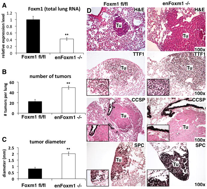
Figure 1. Deletion of Foxm1 from lung endothelial cells increases tumor formation.
A. qRT-PCR analysis of Foxm1 mRNA expression in control Foxm1fl/fl and enFoxm1−/− lungs. RNA was isolated from the total lung. β-actin mRNA was used for normalization. B. Increase in the total number of urethane-induced tumors in enFoxm1−/− mice. enFoxm1−/− and control Foxm1fl/fl mice were administered 6 weekly urethane injections and lungs were harvested 30 weeks after initial urethane injection. C. Increased diameter of tumors in enFoxm1−/− mice. Mean number of tumors per lung (±SE) and mean tumor diameter (±SE) were calculated from n = 12 mouse lungs per group. D. H&E staining demonstrates an increase in the size of lung tumors (Tu) in enFoxm1−/− mice. Tumors in both enFoxm1−/− and control mice are TTF1- and SPC-positive, and CCSP-negative. Magnifications: panels D, 100x; insets, 400x. A p value <0.01 is shown with (**).

