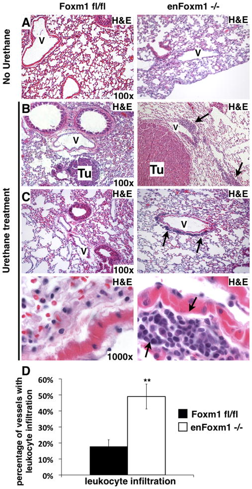
Figure 3. Increased perivascular leukocyte infiltration associates with increased tumor sizes in enFoxm1−/− mice.
A. H&E staining of lungs from enFoxm1−/− and control Foxm1fl/fl mice 30 weeks after urethane treatment. Lung inflammation was not observed in untreated enFoxm1 −/− or control mice. B-C. Increased infiltration of inflammatory cells around bigger tumors (Tu) and vessels in enFoxm1−/− lungs 30 weeks after urethane treatment. Perivascular infiltration of inflammatory cells is shown with arrows. Higher magnification of the representative vessels from control and enFoxm1−/− lungs shown in bottom panels. V – blood vessel. Magnifications: panels A-C, 100x; bottom panels in C, 1000x. D. Percentage of vessels exhibiting leukocyte infiltration was determined in ten random microscope fields and presented as mean + SD.

