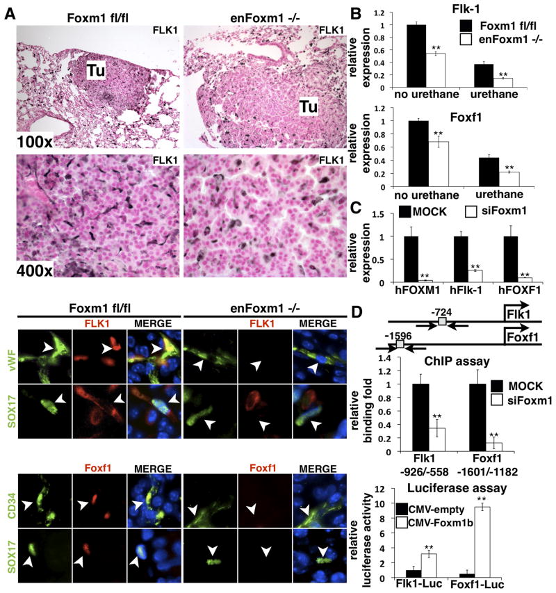
Figure 4. Decreased expression of Flk-1 and Foxf1 in enFoxm1−/− lungs and cultured endothelial cells.
A. enFoxm1−/− mice had decreased Flk1 protein levels in tumors (Tu) compared to control Foxm1fl/fl tumors (top panels). The decrease in Flk-1 and Foxf1 protein levels in enFoxm1−/− tumors was specific to endothelial cells (bottom panels). Immunostaining was performed using antibodies against Flk-1 (red) and either endothelial specific vWF or Sox17 (green). Endothelial specific CD34 or Sox17 (green) antibodies were used to co-localize with Foxf1 (red). The nuclei were counterstained with DAPI (blue). Arrowheads indicate endothelial cells. B. enFoxm1−/− mice showed decreased Flk1 and Foxf1 mRNAs either prior to or after urethane treatment. qRT-PCR was performed using total lung RNA from either untreated mice or mice harvested 30 weeks after urethane treatment. Mouse β-actin mRNA was used for normalization. C. Foxm1 depletion in HMVEC-L cells reduced Flk-1 and Foxf1 mRNA expression. HMVEC-L human endothelial cells were mock transfected (MOCK) or transfected with short interfering RNA (siRNA) duplex specific for Foxm1 mRNA (siFoxm1). Human β-actin mRNA was used for normalization. D. Flk-1 and Foxf1 are direct transcriptional targets of Foxm1. A schematic drawing of promoter regions of the mouse Flk-1 and Foxf1 genes. Locations of potential Foxm1 DNA binding sites are indicated (grey boxes). ChIP assay demonstrated that Foxm1 protein binds to promoter regions of Flk-1 and Foxf1 genes. Foxm1 binding to genomic DNA was normalized to IgG control antibodies. Diminished binding of Foxm1 to the endogenous mouse promoter regions of the Flk-1 and Foxf1 genes was observed after siFoxm1 transfection in MFLM-91U endothelial cells. Luciferase assay demonstrated that Foxm1 induced the transcriptional activity of Flk-1 and Foxf1 promoters. MFLM-91U cells were transfected with CMV-Foxm1b expression vector and luciferase (LUC) reporter driven by either -1.5kb mouse Flk-1 or -2.7kb mouse Foxf1 promoter regions. CMV-empty plasmid was used as a negative control. Cells were harvested at 24 hr after transfection and processed for dual LUC assays to determine LUC activity. Transcriptional activity of the mouse Flk-1 and Foxf1 promoters was increased by CMV-Foxm1b transfection. Magnifications: top panels in A, 100x; middle panels, 400x; bottom panels, 1000x. A p value < 0.05 is shown with asterisk (*).

