
Figure 5.
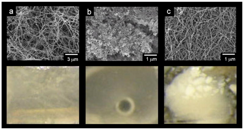
Top, scanning electron micrographs of unmineralized gels of (a) reconstituted collagen, (b) alginate, and (c) peptide amphiphile nanofibers, alginate. All gels were critical point dried to preserve their three-dimensional structure. Bottom, photographs comparing mineral formation after 14 days in a collagen gel, an alginate gel, and a peptide amphiphile gel. The coarse white mineral visible in the PA gel was not observed in collagen or alginate gels.