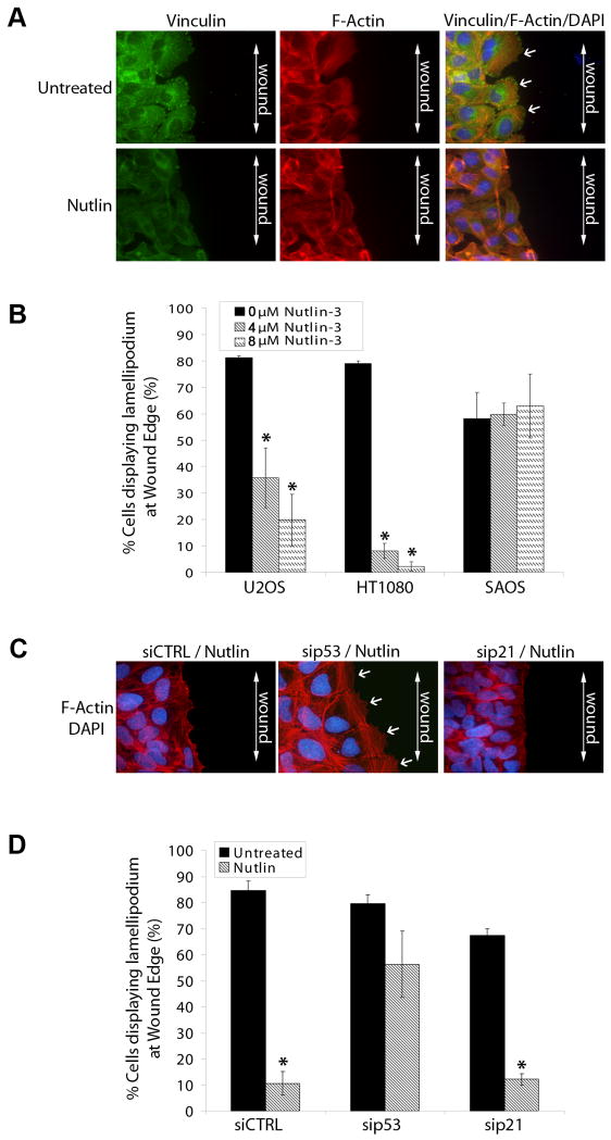
Figure 5. Nutlin decreases lamellipodia formation in wound edge cells in a p53 dependent/p21 independent manner in human cancer cells.
(A) Confluent U2OS, HT1080 and SAOS cells seeded on collagen coated coverslips were treated with Nutlin (4μM/8μM; 40h) or left untreated. Cells were scratched using a sterile micropipette tip and fixed 2h later. (A) Representative fluorescent images of Nutlin treated and untreated U2OS wound edge cells. Cells were stained for focal adhesions (green) and F-Actin stress fibers (red). Nuclei were visualized by DAPI staining (blue). Lamellipodia were identified as flat sheets of F-actin (white arrows) at cell leading edges. Images are representative of three independent experiments each with 200 cells analyzed per experiment.. (B) Percentage of wound edge cells displaying lamellipodia were evaluated and expressed as a percentage of untreated control cells. (* p<0.05 Nutlin treated versus untreated) (C) U2OS cells were transfected with siRNA targeting p21 (sip21), p53 (sip53) or a non targeting control sequence (siCTRL). Cells were treated with Nutlin (8μM) 24h later or left untreated. After 40h, cells were scratched using a sterile micropipette tip and fixed 2h later. Cells were stained for F-Actin stress fibers (red). Nuclei were visualized by DAPI staining (blue). Lamellipodia were identified as flat sheets of F-actin (white arrows) at cell leading edges. Images are representative of three independent experiments each with 200 cells analyzed per experiment.. (D) Percentage of siRNA transfected wound edge cells displaying lamellipodia were evaluated and expressed as a percentage of untreated control cells. (* p<0.05 Nutlin treated versus untreated)

