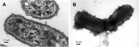
FIGURE 2.
Transmission electron micrographs of vegetative S. cellulosum So ce56 cells grown in liquid medium. A, ultra thin section of vegetative cells clearly shows the presence of two membranes enveloping the bacterial cell. B, negative contrast image of entire So ce56 cells. The cells are surrounded by globular structures representing OMVs.

