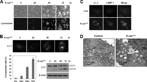
FIGURE 2.
Oncogenic K-Ras induces autophagic vacuole formation in human normal breast epithelial cells. A, MCF10A cells were infected with MFG-control or MFG-K-RasV12. At the indicated times, cells were stained with LysoTracker Green and imaged by fluorescence microscopy. The percentage of vacuolated cells was calculated under fluorescence microscopy. Results from three independent experiments are presented as means ± S.E. (error bars). B, cells were transfected with GFP-LC3 to identify autophagosome and then infected with MFG-control or MFG-K-RasV12. Top, at the indicated times, punctate GFP-LC3 fluorescence was imaged by fluorescence microscopy. Bottom left, the percentage of autophagic cells with punctate GFP-LC3 fluorescence was calculated relative to all GFP-positive cells. Results from three independent experiments are presented as means ± S.E. Bottom right, cell lysates were subjected to immunoblot analysis with anti-LC3 antibody. β-Actin was used as a loading control. C, at 72 h after infection, cells were stained with GFP-LC3 (green) and LAMP-1 (red) to identify the autophagosome. D, representative transmission electron micrographs depicting ultrastructures of MFG-control or MFG-K-RasV12 cells at 72 h. The arrows depict autophagosomes in cells containing recognizable cellular materials.

