FIGURE 5.
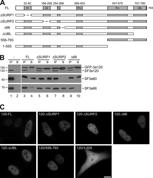
The C terminus of SF3a120 is essential for nuclear import. A, schematic representation of GFP-tagged SF3a120 proteins. Protein domains are as described in the legend to Fig. 1. Dashed lines indicate deleted sequences. B, HeLa lysates from mock-transfected cells or cells transiently transfected with GFP-tagged 3a120-FL, -ΔSURP1, -ΔSURP2, or -Δ66 (as indicated above the panels) were immunoprecipitated with anti-GFP antibodies. Input (IP; 10% of total) and bound (B) proteins were separated by 7.5% SDS-PAGE and detected by Western blotting with anti-SF3a120 (top), anti-SF3a60 (middle), and anti-SF3a66 antibodies (bottom). All immunoprecipitations were performed at the same time, but samples from mock-transfected cells were separated in different gels. GFP-3a120 and the endogenous SF3a subunits are indicated on the right of the panels, the migration of protein markers (in kDa) is shown on the left. C, HeLa cells were transiently transfected with plasmids encoding GFP-tagged SF3a120 proteins as indicated in the individual panels. Fluorescence was monitored in live cells 48 h post-transfection. Bar, 10 μm.

