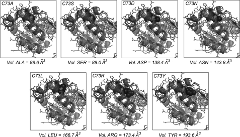
FIGURE 2.
Structural models for the mutations in position 73. Overlay of wild type UROIIIS (Protein Data Bank code 1JR2, light ribbon) and the modeled structure for each Cys-73 mutant (dark ribbon). The side chains for Cys-73 and the specific mutation are represented by red and blue spheres, respectively. Vol., volume.

