
FIGURE 2.
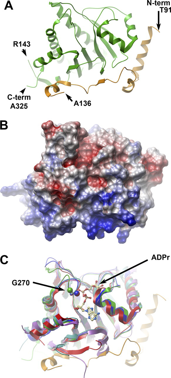
The crystal structure of human MacroD1.A, ribbon representation of the secondary structure of MacroD1. The conserved macrodomain is depicted in green, and the divergent N-terminal (aa 91–136) is in orange. The amino acids at the two termini of the ordered structure are marked, as well as the breakpoints of an internally missing fragment. B, surface potential diagram of MacroD1 shown in the same orientation as Fig. 2A. The N-terminal region (bottom) is highly positive. C, overlay of MacroD1 (green/orange) on the structures of other macrodomains: Feline sarcoma virus (PDB code 3JZT; purple), E. coli YmdB (PDB code 1SPV; blue), human PARP15 (PDB code 3KH6; cyan), and histone macroH2A1.1 (PDB code 3IID; red). An ADPr molecule associated with the Feline sarcoma virus protein is shown as an atomic model. The Cα atom of a conserved glycine (Gly-270 of MacroD1) is marked in a green sphere; the homologous glycine of YmdB and MacroH2A1.1 are marked with blue and red spheres, respectively. The shift of position of the loop containing Gly-270 (green) in MacroD1 relative to all other macrodomain structures is most likely the result of a close contact with a neighboring molecule in the crystal involving residues Val-271 and Phe-272 (not shown).