FIGURE 6.
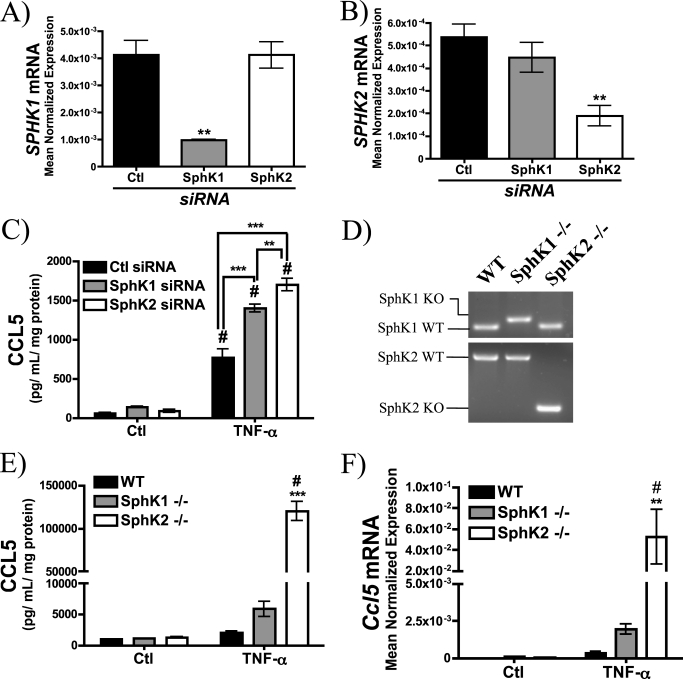
Enhanced up-regulation of CCL5 in cells lacking sphingosine kinase. A, SPHK1, and B, SPHK2 mRNA levels were determined by qRT-PCR after 72 h (20 nm) siRNA treatment (n = 3; one-way ANOVA with Dunnett's post-test; **, p < 0.01). C, CCL5 levels were determined in MCF7 treated with 20 nm control siRNA, SphK1 siRNA, or SphK2 siRNA for 60 h prior to 12 h treatment with PBS (Ctl) or TNF-α (20 ng/ml) (n = 4; two-way ANOVA with Bonferroni post-test; #, p < 0.05 versus untreated cells; **, p < 0.01 versus Ctl siRNA). D, genotyping of WT, SphK1−/−, and SphK2−/− MEFs. E, CCL5 levels were determined in WT, SphK1−/−, and SphK2−/− MEFs in response to 18 h TNF-α (20 ng/ml) (n ≥ 3; two-way ANOVA with Bonferroni post-test; #, p < 0.05 versus untreated cells; ***, p < 0.001 versus WT MEFs). F, Ccl5 mRNA levels in WT, SphK1−/−, and SphK2−/− MEFs in response to 18 h TNF-α (20 ng/ml) were determined by RT-PCR and normalized to β-actin (n ≥ 3; two-way ANOVA with Bonferroni post-test; #, p < 0.05 versus untreated cells; **, p < 0.01 versus WT MEFs).

