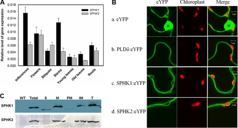
FIGURE 2.
Gene expression and subcellular localization of SPHKs. A, expression of SPHK1 and SPHK2 in Arabidopsis tissues as determined by real-time PCR normalized to UBQ10. RNA was extracted from different tissues of 8-week-old plants. Values are means ± S.E. (n = 3). B, subcellular localization of SPHK1 and SPHK2, using eYFP and PLDδ:eYFP as control. The green color represents eYFP fluorescence and red color marks chloroplasts as a reference. The constructs were transiently transformed into tobacco leaves by infiltration. C, immunoblotting of SPHK1 and SPHK2 in subcellular fractions of Arabidopsis leaves. 25 μg of protein per lane was loaded for total and soluble proteins, and 8 μg for membrane fractions. WT, wild-type total protein from leaves; Total, total protein from transgenic Arabidopsis leaves; S, soluble fraction; M, microsomal fraction; PM, plasma membrane; IM, intracellular membrane; T, tonoplast. SPHK1 was immunoblotted with anti-FLAG antibody, and SPHK2 was immunoblotted with anti-GFP antibody.

