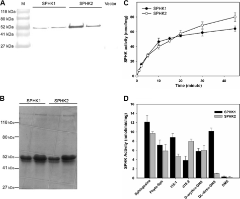
FIGURE 3.
Expression and activity assays of SPHKs. A, immunoblotting of SPHK1 and SPHK2 expressed in E. coli. Total protein (10 μg) was loaded on a SDS-PAGE gel. SPHK1 and SPHK2 were immunoblotted with anti-polyhistidine antibody conjugated with alkaline phosphatase. B, Coomassie Blue staining of purified SPHK1 and SPHK2 from E. coli separated on a 10% SDS-PAGE gel. C, SPHK1 and SPHK2 activity as a function of reaction time. Purified SPHK1 or SPHK2 (3.2 μg) was incubated with 50 μm phytosphingosine for the indicated time. Values are means ± S.E. (n = 3). D, phosphorylation of different LCBs (50 μm) by purified SPHK1 and SPHK2. 0.25 μm enzyme was incubated with substrate for 15 min. Values are means ± S.E. (n = 3).

