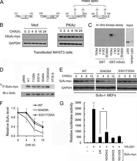
FIGURE 1.
PKA stabilizes Sufu through controlling phosphorylation of Ser-342 and Ser-346. A, Sufu sequences surrounding the four PKA consensus sites. Mass spectrometry (mass spec) analysis shows that Ser-301/Thr-305 and Ser-342/Ser-346 are the actual phosphorylation sites by PKA in vivo. B, Western analysis showing that co-expression of PKAc reduces Sufu turnover after cycloheximide (CHX) treatment. Vect, vector. C, autoradiogram of in vitro kinase assays reveals Sufu as a substrate of PKA but not casein kinase I, casein kinase II, or GSK3β. Input GST and GST-Sufu are shown in the right panel. D, upper panel, autoradiogram of in vitro PKA kinase assay as in C showing that replacing any one of the four PKA consensus sites with alanine is not sufficient to affect Sufu phosphorylation but replacing all four sites abolished it. Lower panel, Western analysis of Myc-Sufu and mutants expressed by in vitro translation. IB, immunoblot. Western blot analysis (E) and quantification (F) of the turnover rate of Sufu and its mutants in transfected Sufu−/− MEFs. The data presented in F were derived from three repeated experiments. G, luciferase reporter assay for various Sufu mutants co-transfected with Gli1 and the 8×GliBS construct in Sufu−/− cells with Renilla luciferase as an internal control. Each data point represents results from triplicate wells. Error bars are standard deviations. CHX, cycloheximide. *, p < 0.01.

