FIGURE 5.
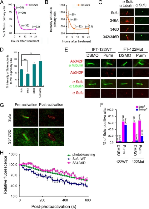
Phosphorylation promotes ciliary retention of Sufu. In Ptch−/− cells, immunofluorescent staining indicated that KT5720 treatment led to a rapid decrease of the percentage of Sufu-positive primary cilia (A) and the intensity of Sufu in primary cilia (B). Sufu was typically found in ∼95% of cilia, but this number decreased to ∼65% after KT5720 treatment for 24 h. C, representative autofluorescence images of Sufu-GFP and Sufu mutants in transfected Sufu−/− cells. Replacing Ser-346 with alanine decreased while replacing Ser-346 or both Ser-342 and Ser-346 with aspartic acid ciliary localization. Cilia were visualized by staining with anti-acetylated α-tubulin (red). The number of cilia counted for each data point was between 17 and 24. D, quantification of C. *, p < 0.01; **, p < 0.001. E, representative immunofluorescent staining of phospho-specific and total Sufu at ciliary tips in wild type or IFT122 null MEFs. Purmorphamine (Purm) treatment induced immunofluorescent staining of p-Sufu and total Sufu at the tips of primary cilia. In IFT122 null MEFs, p-sufu was not detected whereas total Sufu accumulated at the tip of cilia with or without purmorphamine treatment. F, quantification of E. G, live cell imaging of photoactivatable Sufu and S342D/S346D mutant force-expressed from PA-mCherry1-N1-Sufu and PA-mCherry1-Sufu-S342D/S346D, respectively, in Ptch−/− cells. Somatostatin receptor-3-GFP was co-transfected to mark for cilia. A type area to be photoactivated was marked, and images of merged green and red channels taken pre- and post-photoactivation were shown. H, relative fluorescence of PA-mCherry-Sufu or PA-mCherry1-Sufu-S342D/S346D was calculated at each time point after photoactivation by correcting for photoextinction of mCherry fluorescence from the total intensity recorded in the red channel at ciliary tips.

