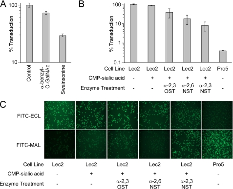
FIGURE 3.
A, effect of glycosylation inhibitors on AAV9 transduction. Prior to infection with AAV9 vectors (m.o.i. = 1000 vg/cell), CHO Lec2 cells were treated with α-benzyl-GalNAc (1 μg/ml) or Swainsonine (10 μm), inhibitors of O- and N-glycosylation, respectively. B, effect of enzymatic resialylation on AAV9 transduction. CHO Lec2 cells were treated with 50 milliunits/ml each of α2,3-(N)-sialyltransferase (α2,3NST), α2,6-(N)-sialyltransferase (α2,6NST), or α2,3-(O)-sialyltransferase (α2,3OST) and 1 mm CMP-sialic acid (Sigma) to resialylate cell surface asialo glycans. Luciferase transgene expression was quantified at 24 h after infection and is expressed as percent infectivity with respect to untreated or wild type (CHO Pro5) control. All experiments were carried out in triplicate. Error bars indicate S.E. C, lectin staining of CHO cell lines subjected to enzymatic resialylation. CHO Lec2 cells treated with CMP-sialic acid alone or with CMP-sialic acid and different sialyltransferases were subjected to lectin staining using FITC-labeled ECL, which exclusively recognizes Gal(β1,4)GlcNAc or FITC-labeled MAL, which recognizes α2,3-sialylated Gal(β1,4)GlcNAc. Untreated wild-type CHO Pro5 cells, which show high levels of FITC-MAL I staining and untreated Lec2 cells, which show high levels of FITC-ECL staining, were included as control.

