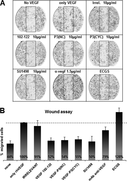
FIGURE 7.
HUVEC migration in scratch wound assay using peptides as inhibitors. A, pictures taken at ×40 magnification in light microscopy. B, average percentage of migrated cells, counted using the software ImageJ (National Institutes of Health) assuming rhVEGF control as 100%. Inhibitors were used at indicated concentration. α-vegf represents a monoclonal antibody demonstrated to block VEGF-dependent pathways. Results shown represent an average of two different experiments each performed in duplicate, and error bars represent mean ± S.E. IRR, irrelevant.

