
FIGURE 3.
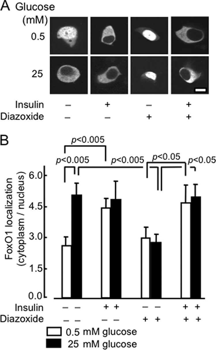
Role of secreted insulin on the effects of glucose on FoxO1-EGFP translocation. A, after 24 h of transient transfection with pFoxO1-EGFP, MIN6 cells were incubated at varying concentrations of insulin (0, 20 nm), glucose (0.5, 25 mm), or DAO (diazoxide, 20 and 200 μm) for 1 h in medium prior to imaging. B, distribution of FoxO1 between the cytosol and nucleus. Data show ratio (mean ± S.E., n = 20–50 cells from three separate experiments).