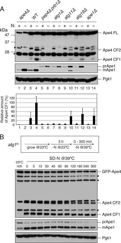
FIGURE 4.
Ape4 undergoes limited cleavage in vacuolar proteolysis- and autophagy-dependent manner. A, the ape4Δ (YMY111; lanes 1 and 2), wild-type (WT; SEY6210; lanes 3 and 4), pep4Δ prb1Δ (YMY105; lanes 5 and 6), atg1Δ (WHY1; lanes 7 and 8), atg11Δ (YTS147; lanes 9 and 10), atg19Δ (SSY31; lanes 11 and 12), and ape1Δ (YMY103; lanes 13 and 14) cells were grown in YPD medium (lanes 1, 3, 5, 7, 9, 11, and 13) and starved in SD(-N) medium for 3 h (lanes 2, 4, 6, 8, 10, 12, and 14). The cell extracts (0.1 A600 nm cell equivalent) were analyzed by immunoblot probed with anti-Ape4 (upper panel) and anti-Ape1 (middle panel) antisera and anti-Pgk1 (lower panel) antibody. A single dot and double dot indicate CF1 and CF2, respectively. Quantification of CF1 and Pgk1 were done using ImageJ software. The band intensity of CF1 was normalized by that of Pgk1 and plotted relative to WT treated with SD(-N). Error bars indicate the S.D. of three independent experiments. B, the ape4Δ atg1Δ (YMY112) cells were transformed with an atg1ts plasmid (pAtg1ts) and a plasmid expressing GFP-Ape4 from the CUP1 promoter (pTS555). The strain was grown in SC-Ura-Trp medium and starved in SD(-N) at a permissive temperature of 23 °C for 3 h. After shifting to the nonpermissive temperature of 39 °C, samples were collected at the indicated time points and analyzed by immunoblot using anti-Ape1 (upper panel) and anti-Ape4 (middle panel) antisera and anti-Pgk1 (lower panel) antibody. FL, full length. Arrowheads indicate nonspecific proteins.

