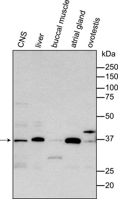
FIGURE 2.
Western analysis of A. californica tissue expression of DAR1. Tissue protein lysates of CNS, liver, buccal muscle, atrial gland, and ovotestis tissues were subjected to reducing SDS-PAGE. Ten micrograms of each sample were co-run with Precision Plus Protein Standards in 12% Tris-glycine gel. The transfer blot was stained with rabbit anti-DAR1 serum (1:30,000) and goat anti-rabbit HRP conjugate (1:20,000). Film was exposed for 5 min. The predicted native DAR1 protein is 35.4 kDa.

