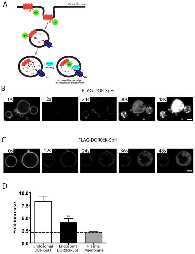
Figure 4. Intralumenal wild -type and lysyl-mutant receptors visualized using a pH-sensitive GFP variant.
A) Schematic of experimental setup following receptors fused to the GFP variant (ecliptic pHluorin) which fluoresce when located in the cytoplasm but whose fluorescence is efficiently quenched when exposed to the acidic environment of the endosome lumen. Addition of the weak base chloroquine neutralizes endosomal pH and reveals any tagged receptors present in the endosome lumen. B and C) HEK293 cells were transiently transfected with CFP-Rab5Q79L and either F-DOR-SpH (B) or F-DOR-0cK-SpH (C), and replated onto to coverslips before treatment for 90 minutes with 10μM DADLE. Cells were then imaged at a rate of 5 frames per second by spinning disc confocal microscopy and treated with 1mM chloroquine after 50 frames. Image sequences are shown for cells expressing F-DOR-SpH (B) or F-DOR-0cK-SpH (C), see Supplemental Movies 3 and 4 for full sequence. D) Quantification of the chloroquine-induced increase in fluorescence intensity. Mean and SEM are shown for F-DOR-SpH (Endosomal DOR-SpH, n= 8 cells, 24 endosomes) and F-DOR0cK-SpH containing endosomes (Endosomal DOR0cK-SpH, unpaired t-test; ***, p<0.01, n= 7 cells, 22 endosomes), and F-DOR-SpH expressed on the plasma membrane (Plasma Membrane, n= 11 cells). Scale bar = 2 μm.

