Figure 2.
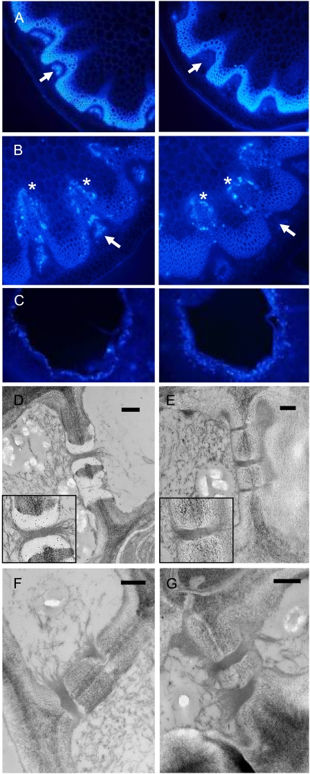
Loss of callose from the phloem in flowering stems of the gsl7 mutant. All images are representative of results obtained from several different wild-type and gsl7 mutant plants. The cell walls of the xylem and fibers are autofluorescent. A, The basal 1 to 2 cm of flowering stems from 45-d-old plants was excised, and hand-cut sections were stained with aniline blue and viewed with a UV epifluorescence microscope. Left, wild-type plant; right, gsl7 mutant plant. Arrows indicate the position of the phloem. Fluorescence indicating the presence of callose is associated with the phloem in the wild type but not in the gsl7 mutant. B, As in A, but excised stems were incubated in water for 18 h prior to staining. Left, wild-type plant; right, gsl7 mutant plant. Fluorescence indicating the presence of callose is associated with cells adjacent to the xylem in both wild-type and gsl7 mutant stems (asterisks) but is associated with the phloem only in wild-type stems (arrows). C, Fully expanded leaves from 42-d-old plants were punctured with a plastic tip and then harvested the next day, stained with aniline blue, and viewed with a UV epifluorescence microscope. Left, wild-type plant; right, gsl7 mutant plant. D to G, Electron micrographs of phloem elements in sections of the basal 1 to 2 cm of flowering stems from wild-type (D) and gsl7 mutant (E–G) 45-d-old plants excised directly into glutaraldehyde fixative. Sections in D and E are immunogold labeled with an anti-callose antiserum. Note the presence of gold particles in the electron-transparent sieve plate lining of wild-type plants and their absence from an equivalent position in mutant plants (compare enlarged insets in D and E). Bars = 500 nm.

