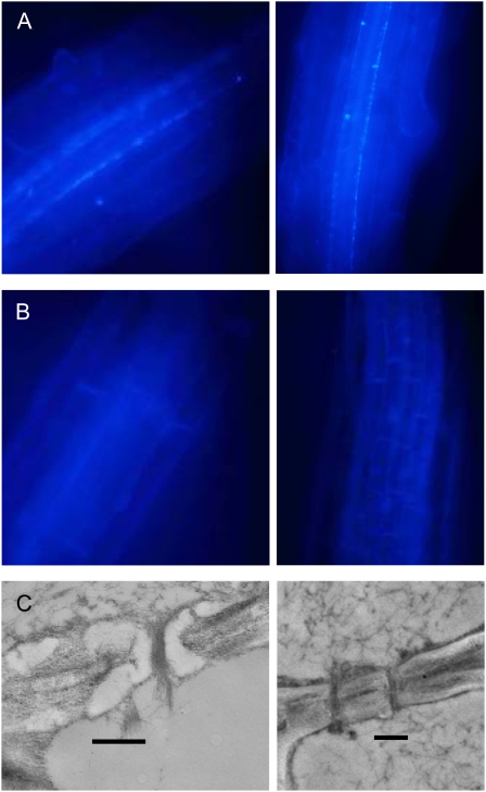
Figure 3.
Loss of callose from the phloem in roots of the gsl7 mutant. All images are representative of results obtained from several different wild-type and gsl7 mutant plants. Photographs are of roots from 4-d-old seedlings. A and B, Regions behind the expansion zones of roots from two different seedlings stained with aniline blue. In wild-type seedlings (A), fluorescence is associated with two cell files in the stele. In gsl7 seedlings (B), no fluorescence is visible in these cell files. C, Electron micrographs of phloem elements in sections of the proximal region of roots of wild-type (left) and gsl7 mutant (right) seedlings. An electron-transparent lining is present in the sieve plate pores of wild-type but not gsl7 plants. Bars = 500 nm.

