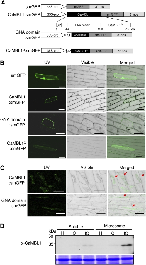
Figure 3.
Subcellular localization of CaMBL1. A, Schematic representation of CaMBL1 constructs. B, Bright-field images of subcellular localization of CaMBL1:smGFP, GNA domain:smGFP, and CaMBL1C:smGFP proteins in onion epidermal cells. GFP images were observed by confocal laser scanning fluorescence microscopy. Bars = 0.1 mm. C, Subcellular localization of CaMBL1:smGFP and GNA domain:smGFP in onion epidermal cells after plasmolysis with 1 m NaCl. Red arrows indicate plasmolyzed plasma membrane. Bars = 0.1 mm. D, Immunodetection of CaMBL1 with anti-CaMBL1 antibody in soluble and microsomal fractions from pepper leaves infected by Xcv. H, Healthy leaves; C, compatible; IC, incompatible.

