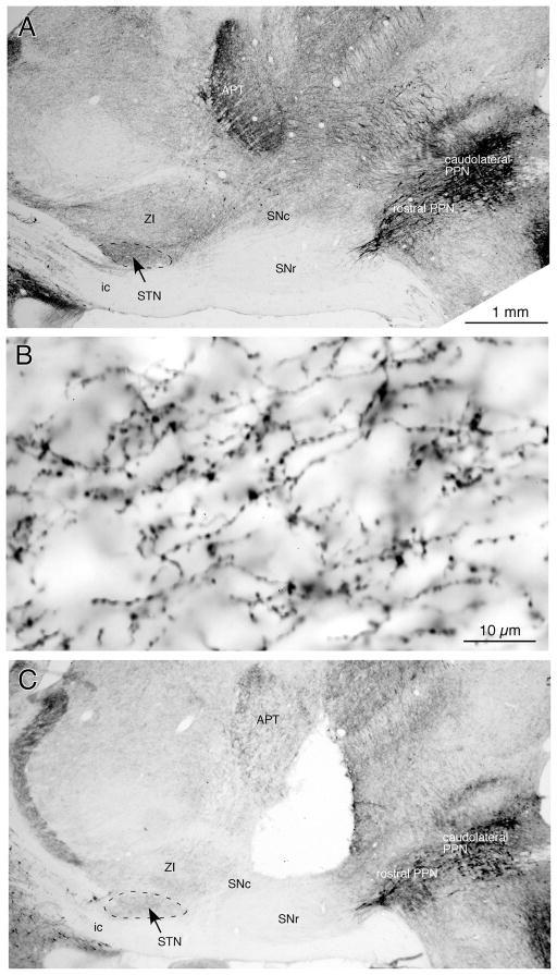
Fig. 1.
Photomicrographs of sagittal sections immunostained for ChAT. A: A low magnification view shows a large number of ChAT+ neurons in the PPN. The STN contains a dense plexus of ChAT+ fibers, and ZI contains a moderate number of ChAT+ fibers. The SNc also contains a moderate number of ChAT+ fibers, while the SNr contains very few. B: A high magnification view of ChAT+ fibers in the STN. Thin ChAT+ fibers with numerous small boutons en-passant run from the caudodorsal to rostroventral direction. C: Electrical lesions of the midbrain tegmentum extending from the deep layers of the superior colliculus to the SNc greatly reduced the ChAT+ fibers in the STN and ZI. See Abbreviations list for the abbreviations used in this and subsequent figures.

