Fig. 6.
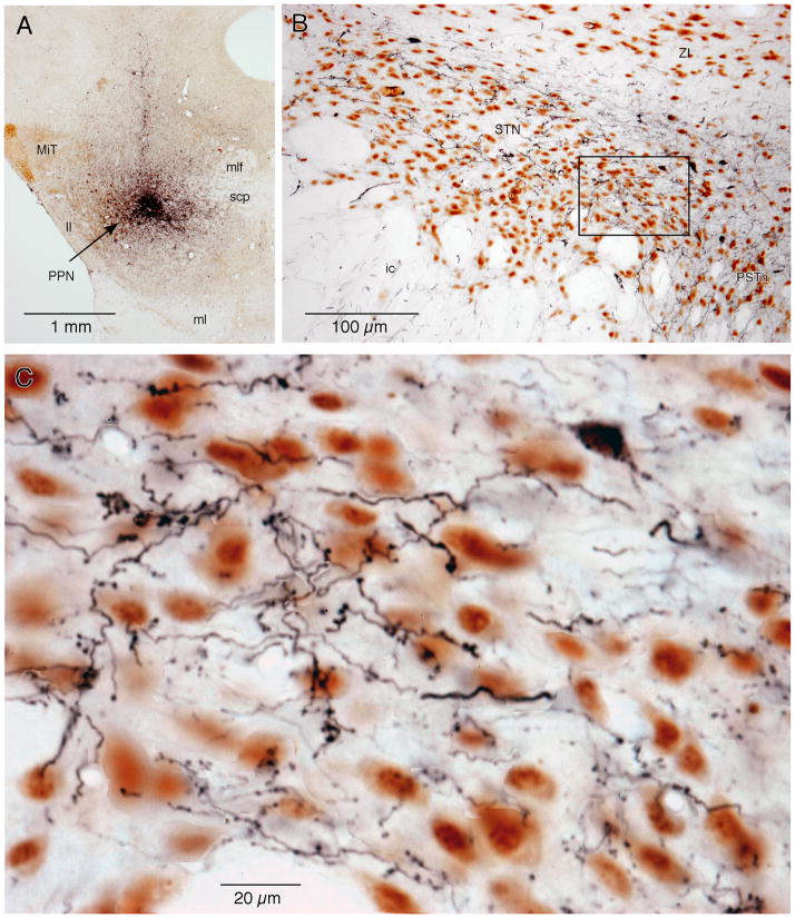
Photomicrographs show BDA-labeled fibers in the STN after BDA injection into the PPN. A: A section doubly stained for BDA and ChAT. The BDA injection site was dorsomedial to the PPN. B: A section doubly stained for BDA and NeuN. BDA-labeled fibers can be seen in the STN, ZI, ic, and PSTh. C: A high magnification view of the area marked by the box in B. Two types of fibers, thin fibers with small en passant varicosities and slightly thicker fibers with clumps of larger boutons, can be seen in the STN. The area also contains a retrograde-labeled STN neuron.

