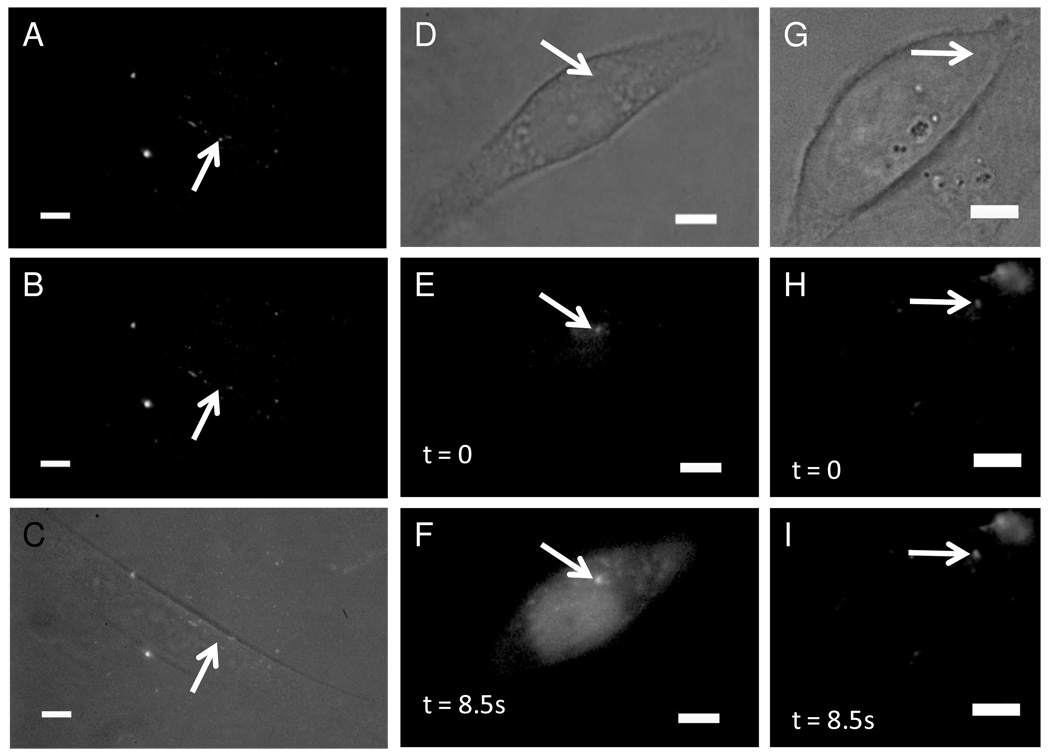
Figure 2. Release of encapsulated contents from endocytosed lipid nanocapsules into the cytoplasm.
The lipid nanocapsules were photolyzed by a single 3-ns pulse of 645-nm light after 2 hours of incubation followed by 2 washing steps. (A–C) CHO-M1 cells loaded with 100-nm-diameter nanocapsules consisting of 9:1 DPPC:DOPC+4mol% DiD and 50µM Alexa-488. (D–F) CHO-M1 cells loaded with the calcium indicator dye fluo-3 and 100-nm-diameter nanocapsules made of 9:1 DPPC:DOPC+4mol% DiD+1mol% DiO and 730µM IP3. (G–I) CHO-M1 cells loaded with fluo-3 and empty 100-nm-diameter nanocapsules made of 9:1 DPPC:DOPC+4mol% DiD+1mol% DiO. Fluorescence is from 488nm excitation in the epifluorescence configuration; the scale bar is 5µm.

