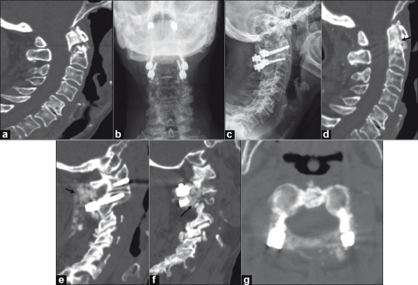
Figure 4.

A 78-year-old woman with nonunion of a Type II odontoid fracture. Preoperative CT imaging with sagittal reconstruction demonstrates a Type II odontoid fracture (panel a). Postoperative plain AP (panel b) and lateral (panel c) radiographs following C1-C2 instrumented arthrodesis with C1 lateral mass and C2 pedicle screws, rhBMP-2 and cancellous allograft. CT imaging with sagittal reconstructions at one-year follow-up demonstrates healing of the odonoid fracture (panel d, arrow) and brisk bony fusion (panels E and f, arrows). Axial CT imaging at the interspace of C1-C2 does not demonstrate canal encroachment (panel g).
