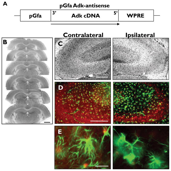
Figure 3.
pGfa- Adk-antisense virus selectively knocks down ADK expression in astrocytes. (A) Schematic illustration of the pGfa-Adk-antisense (Adk-AS) vector coding region. The Adk-cDNA is oriented in the antisense direction (3′ to 5′) under control of the truncated astrocyte specific gfaABC1D promoter (pGfa). The transcriptional woodchuck hepatitis virus transcriptional regulatory element (WPRE) is placed downstream of the Adk-cDNA to induce expression of intronless viral messages and increase the stability and level of gene expression. (B-E) Coronal sections of wild-type mice subjected to unilateral injection of Adk-AS virus into the CA3 region of the hippocampus. (B, C) ADK immunohistochemistry with ADK primary antibodies and DAB enhancement. (B) Unilateral injection of Adk-AS causes localized knockdown of ADK in CA3 (black box). (C) Higher magnification image shows that Adk-AS decreases astrocytic ADK expression ipsilateral to the injection site (right panels) compared to the non-injected contralateral side (left panels). (D, E) Dual immunofluorescence analysis using ADK (red) and GFAP (green) primary antibodies. (D) The decreased ADK immunoreactive material in the ipsilateral hippocampus (right panel) is confined to the CA3. (E) Higher magnification image shows that ADK is uniformly knocked down within GFAP-positive astrocytes (right panel). Scale bars = 1 mm B, 250 μm B, C; 10 μm D.

