. 2009 Sep;11(3):269–280. doi: 10.31887/DCNS.2009.11.3/dalewis

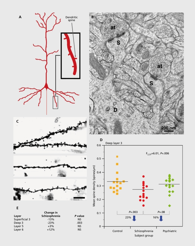
Figure 2. Pyramidal neuron dendritic spines in the human DLPFC. A) Schematic diagram illustrating the dendritic tree and dendritic spines on a prototypic pyramidal neuron. B) Electron micrograph showing a dendrite (D) with two spines (S). Each spine receives an asymmetric (presumably excitatory) synapse from an axon terminal (at). C) Golgi-impregnated basilar dendrites and spines on deep layer 3 pyramidal neurons from a normal comparison (top) and two subjects with schizophrenia (bottom). Note the reduced density of spines in the subjects with schizophrenia in these extreme examples. D) Scatter plot demonstrating the lower density of spines on the basilar dendrites of deep layer 3 pyramidal neurons in the DLPFC of subjects with schizophrenia relative to both normal and psychiatrically-ill comparison subjects. E) Laminar-specificity of the spine density differences in the same subjects. Reproduced from ref 1: Lewis DA, Gonzalez-Burgos G. Neuroplasticity of neocortical circuits in schizophrenia. Neuropsychopharmacology. 2008;33:141-165. Copyright© Nature Publishing Group 2008.

Figure 2.