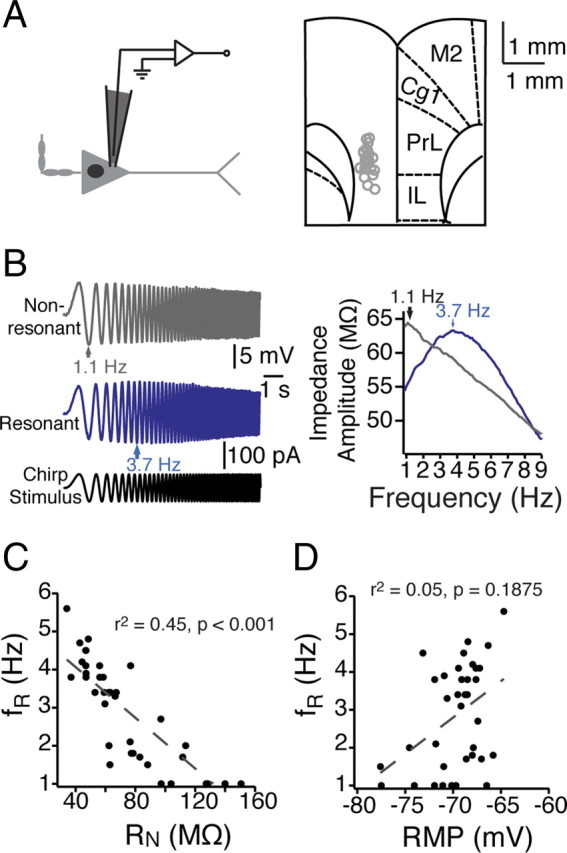
Figure 1.

Heterogeneity in the dynamic properties of mPFC neurons. A, Somatic recordings of layer V mPFC neurons were conducted within ventral mPFC, including prelimbic and infralimbic cortex. Left, A schematic of an mPFC neuron and the somatic recording location. Right, Recording locations overlaid with a modified version of a coronal diagram from a rat brain atlas (Paxinos and Watson, 1993). B, In response to a 10 s chirp stimulus at their resting membrane potential, different mPFC neurons resonated across a range (1–6 Hz) of frequencies. C, D, In addition to exhibiting different resonance profiles, neurons were diverse in both steady-state input resistance (C) and resting membrane potential (D) (n = 38 neurons). Gray dashed lines represent the linear fit of the data, with correlation values listed. PL, Prelimbic; IL, infralimbic; Cg1, anterior cingulate; M2, secondary motor cortices.
