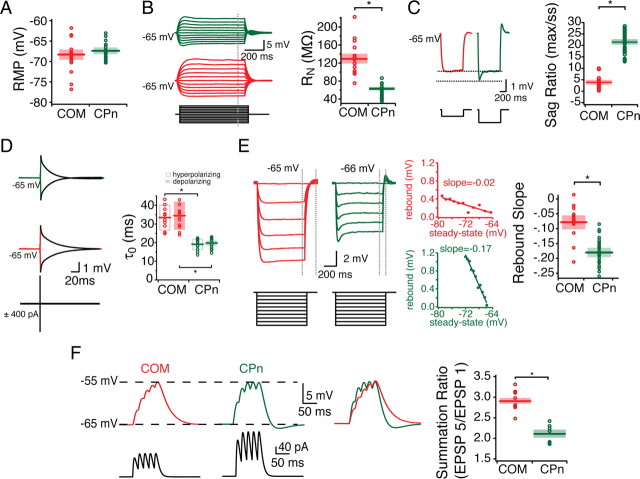
Figure 4.
Subthreshold properties of CPn and COM neurons. Somatic patch recordings of labeled COM and CPn neurons reveal that they have distinct subthreshold properties. A, CPn neurons are slightly more depolarized than COM neurons, although not significantly. For the purpose of comparison, neurons were held at −65 mV during stimulus protocols. B–F, COM neurons (red) have higher steady-state input resistance (B), lower sag ratio (C), slower time constant in both the hyperpolarizing and depolarizing directions (D), less rebound (E), and more temporal summation (F) than CPn neurons (green). *p < 0.05.

