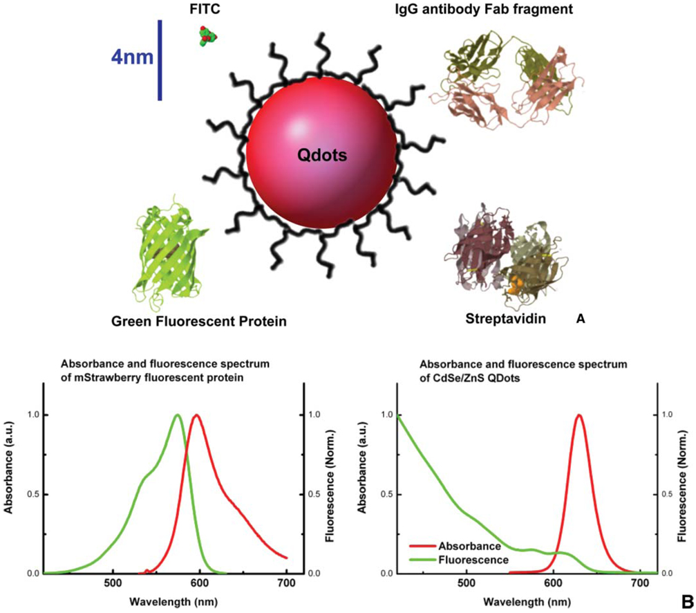
Figure 1.
(online colour at: www.biophotonics-journal.org) (A) Comparison of fluorophores and commonly used targeting proteins shown to scale. While Qdots as synthesized core/shell structure have diameters ranging from 2–10 nm, the solubilization interface and biological functionality coating significantly increase the size of the final products. Here a CdSe/ZnS quantum dot coated with peptides (Qdot in red, and peptide coating in black) is shown to compare with FITC fluorophore, green fluorescent protein, IgG Fab fragment, and streptavidin. The hydrodynamic diameter of peptide-coated red-emitting CdSe/ZnS quantum dots is ~12 nm [45]. Streptavidin coated Qdots usually end up with diameters ~15–20 nm, and antibody Fab fragments coupled Qdots can be as large as 25 nm in diameter. Scale bar, 4 nm. (B) Comparison of absorption and fluorescence spectra of FPs to Qdots. Normalized absorbance is in green, and normalized fluorescence emission is in red. The spectra of one red emitting fluorescent protein mStrawberry [71] is shown on the left, and the spectra of red emitting CdSe/ZnS Qdot is shown on the right. The absorption band of typical organic fluorophores is narrow, and the emission peak is broadened on the red shoulder. Qdots, on the contrary, have a broadband absorption spectrum and symmetric, narrow emission peak.

