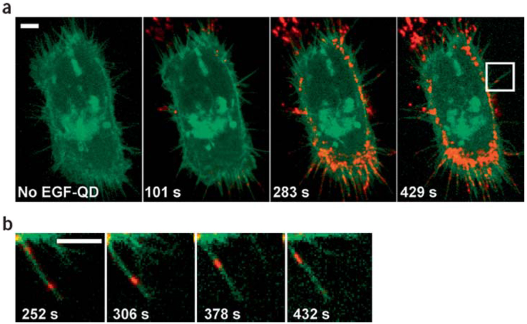
Figure 4.
(online colour at: www.biophotonics-journal.org) Retrograde transport of EGF conjugated Qdots along filopodia. (a) A431 cells expressing endogeneous erbB1 and erbB3-eGFP (in green) were labeled with Qdots (in red) conjugated to epidermal growth factor (EGF), which binds to the transmembrane receptor erbB1. Confocal images at different time points were shown in series, indicating the endocytosis of Qdots-ligand bound receptors. (b) A single filopodium of the cell indicated in (a) was magnified, demonstrating the migration of EGF-Qdots towards the cell body at a velocity ~10 nm/sec. This figure was reprinted by permission from Macmillan Publishers Ltd: Nature Biotechnology [53], copyright 2004.

