Fig. 3.
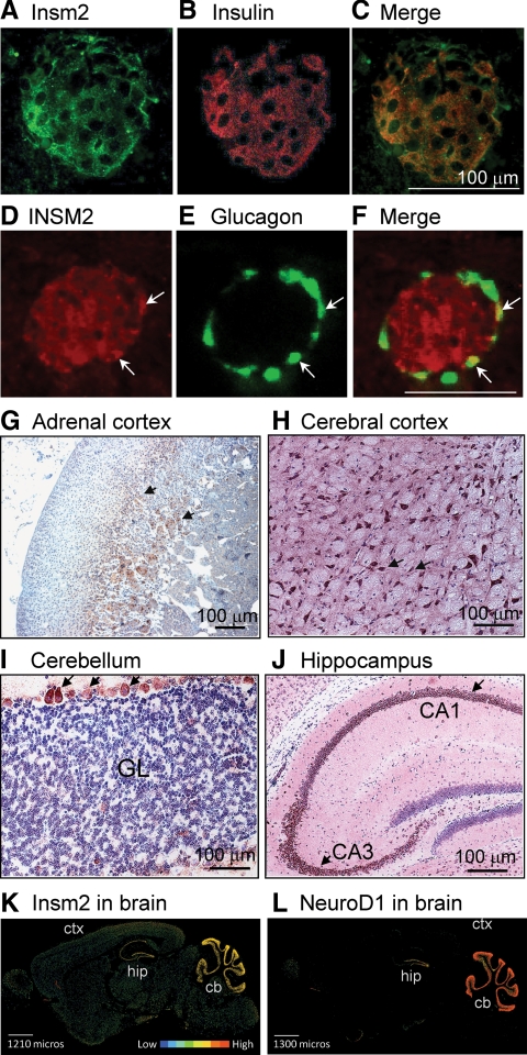
INSM2 in neuroendocrine tissues. A–C, INSM2 was expressed in mouse pancreatic islet β-cells and colocalized with insulin. D–F, INSM2 also was detected in human islet α-cells because it was colocalized with glucagon (arrows). G, By histochemistry stainings, INSM2 was found in the deeper layers of the adrenal glands (arrows). INSM2 also was expressed in neuronal cells of the cerebral cortex (H), Purkinje cells of the cerebellum [granule layer (GL)], (I), and the hippocampus (J). K and L, Representative images of in situ hybridization in the adult mouse brain (sagittal). mRNAs of Insm2 (K) and NeuroD1 (L) are detected with a similar pattern in cerebellum (cb), hippocampal region (hip), and to a much lesser extent in cerebral cortex (ctx). Expression intensities are shown in color bars.

